


Description

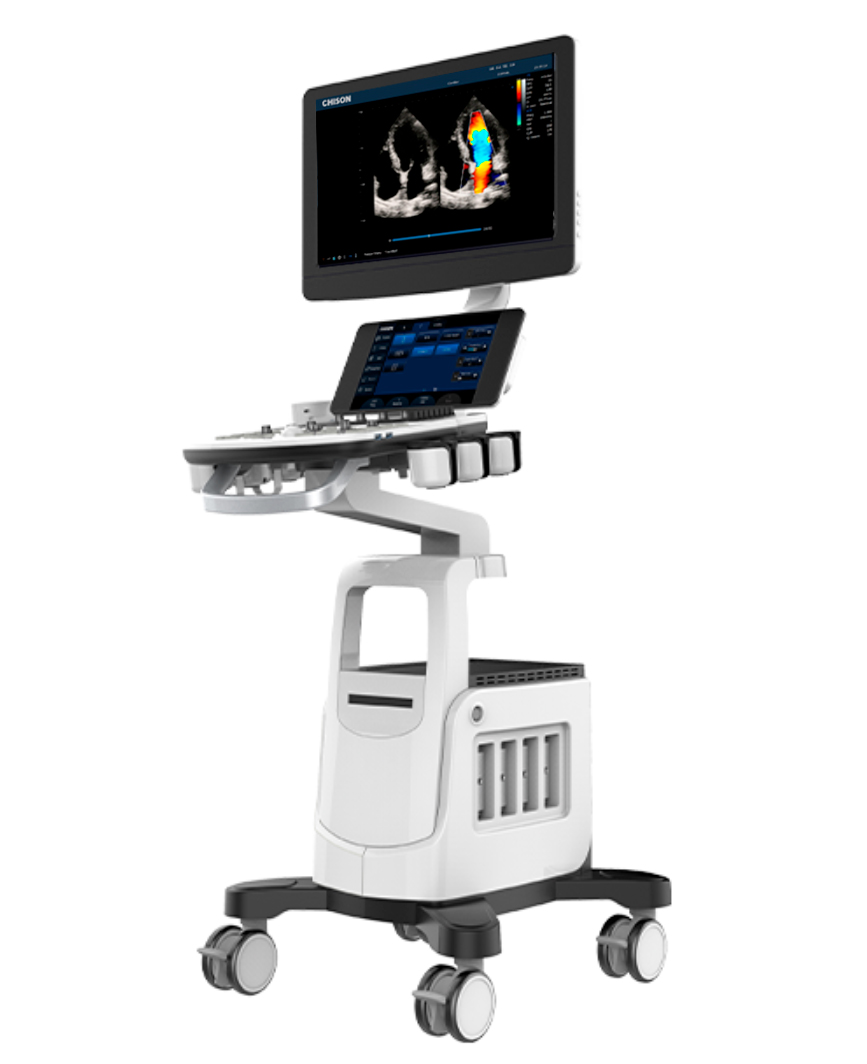
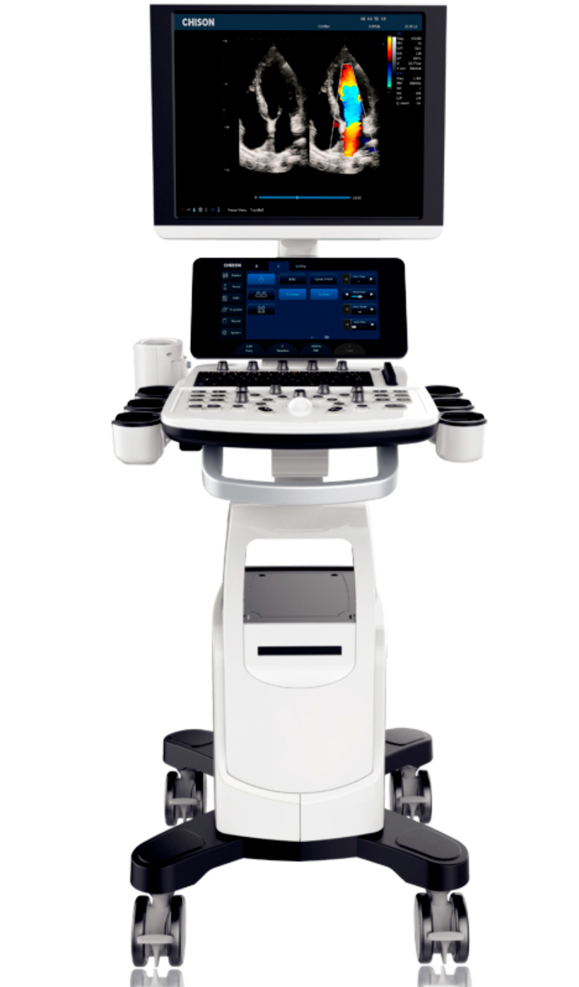
Chison CBit 4 Ultrasound, ideal for providing health professionals with exceptional diagnostic capabilities.
With this equipment, you can experience an intuitive workflow and intelligent features that optimize efficiency and precision in diagnosis. The CBit series offers competitive pricing options for medical consultations of all needs.
The standard configuration includes:
- Main unit with high resolution 19" LCD monitor + 10.1" touch screen + 3 probe connectors.
- 500G hard drive.
- 6 USB ports.
- 7.0 Mhz linear probe.
- 4D module + CW module.
- TGC and LGC.
- B, 2B, 4B, B/M, B/BC, CFM, PW, Power Doppler/PD directional, Instant Triplex, Duplex, Quadplex, Trapezoidal, Chroma B&M&PW, Full screen.
- Real-time automatic PW tracking and measurement.
- Super Image Module: FHI, Multiple composite images, SRA, AIO.
- Q-Image (intelligent image optimization), X-contrast, Q-beam, Q-Flow.
- Measurement and calculation software packages: general, obstetrics and gynecology, cardiac
- 2D Steer, HIPPA.
Features:
- Intelligent focus: Automatically detects the focus position based on depth and focuses on the study area to improve quality.
- Raw data: Allows you to adjust parameters after freezing. Speeds up scan time and saves processing time.
- Wide-angle TV probe: Provides more diagnostic information with an extremely wide angle of up to 210°, saving time and improving efficiency.
- Intelligent volume split: Allows 3D cuts to be made for meticulous observation in the tomographic view. Especially indicated for deep diagnoses, such as cleft lip.
- HD Niche: Allows user to view sagittal, axial and coronal view of a target by moving the cross point of the three orthogonal planes, helping the user to better understand the internal shape and structure of the study object.
- Intelligent HIP: Use the chart for orthosis hip diagnosis. Different angles indicate different levels of hip deformity, which is easier to see with the help of this chart. (I, II, D, IIIa, IIIb).
- HD Zoom: Allows color information to be enlarged while maintaining high resolution. It is important for detecting blood information from small vessels, especially for fetal heart diagnosis.
- Virtual Convex: Allows to enlarge the scanning area in the convex probe, just like the convex trapezoid. It is especially indicated for the display of large organs, especially liver or kidney through the costal space.
Fields of application, depending on the probe used:
- Obstetrics.
- Abdomen.
- Vascular.
- Cardiology.
- OB/Gynecology.
- Urology.
- Musculoskeletal.
- Soft tissues/small body parts.
- Anesthesia.
- Pediatrics.
- Orthopedics.
Alternative products


4D convex probe 2.0-6.8 MHz for ultrasound machines of the CBit and...
6.390,00€(7.731,90€ VAT INCLUDED)
Linear probe 4.0-15.0 MHz for ultrasound machines from the CBit and...
2.190,00€(2.649,90€ VAT INCLUDED)

Linear probe 4.0-15.0 MHz for ultrasound machines from the CBit and...
6.235,00€(7.544,35€ VAT INCLUDED)